When You Think Heart Attack Prevention, Think Cleerly
Cleerly’s AI-enabled CCTA solution provides comprehensive, accurate, and trackable insights to personalize heart attack prevention
Request a demoNot All Plaque and Plaque Analysis Software Are the Same
ACC/AHA guidelines highlight the importance of plaque, particularly high-risk plaque, as a driver of major adverse cardiovascular events (MACE)1
Assessing atherosclerosis is challenging due to limitations of current image processing4
Cleerly Advanced Coronary Plaque Analysis
Predicting cardiovascular risk with AI precision
Vessel-by-Vessel Detail
Precision phenotyping for each artery and branch with stenosis quantification and vascular remodeling scores
Comprehensive Plaque Assessment
At-a-glance view of characterized plaque volume by coronary region
Stenosis Scoring
Clear and concise summary of identified stenoses by severity
Supports Next-Gen Hardware
Advanced segmentation on photon-counting CT
Level 3 Expert Cleerly was superior compared with Level 3 Expert Readers in determining stenosis severity, plaque volume, and composition.7 |
Quantitative Coronary Angiography (QCA) Cleerly demonstrated high diagnostic performance for determining stenosis severity.9 |
Myocardial Perfusion Imaging Cleerly demonstrated higher diagnostic performance than MPI for detecting obstructive coronary artery disease.11 |
Intravascular Ultrasound Cleerly demonstrated high agreement with IVUS for absolute measures of coronary lumen size to determine accurate stenoses measurements.10 |
Near-field Infrared Spectroscopy (NIRS) Cleerly demonstrated high agreement with NIRS for the identification of lipid-rich plaques.10 |
Backed by Clinical Evidence
See the demonstrated clinical evidence of the Cleerly AI platform
Earlier Plaque Detection15
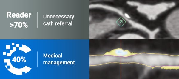
In the CONSERVE trial
(N=747)
53%
less significant stenoses than clinical readers,
leading to less unnecessary downstream testing
In a retrospective analysis of the PARADIGM trial
(N=99)
87%
of plaques were present at the same location
as small plaques on baseline CCTA
References:
1 Gulati M, Levy PD, Mukherjee D, et al. Circulation. 2021;144(22):e368-e454. doi:10.1161/CIR.0000000000001029
2 Chang HJ, Lin FY, Lee SE, et al. J Am Coll Cardiol. 2018;71(22):2511-2522. doi:10.1016/j.jacc.2018.02.079
3 Nurmohamed NS, Cole JH, Budoff MJ, et al. Eur Heart J Cardiovasc Imaging. 2024;25(6):857-866. doi:10.1093/ehjci/jeae029
4 Nieman K, García-García HM, Hideo-Kajita A, et al. J Cardiovasc Comput Tomogr. 2024;18(5):429-443. doi:10.1016/j.jcct.2024.05.232
5 Nurmohamed NS, Danad I, Jukema RA, et al. JACC Cardiovasc Imaging. 2024;17(8):894-906. doi:10.1016/j.jcmg.2024.01.007
6 Karlsberg RP, Nurmohamed NS, Quesada CG, et al. Int J Cardiol Heart Vasc. 2024;53:101433. doi:10.1016/j.ijcha.2024.101433
7 Choi AD, Marques H, Kumar V, et al. J Cardiovasc Comput Tomogr. 2021;15(6):470-476. doi:10.1016/j.jcct.2021.05.004
8 Hakim D, Coskun AU, Maynard C, et al. J Cardiovasc Comput Tomogr. 2023;17(3):201-210. doi:10.1016/j.jcct.2023.03.009
9 Griffin WF, Choi AD, Riess JS, et al. JACC Cardiovasc Imaging. 2023;16(2):193-205. doi:10.1016/j.jcmg.2021.10.020
10 Omori H, Matsuo H, Fujimoto S, et al. Atherosclerosis. 2023;386:117363. doi:10.1016/j.atherosclerosis.2023.117363
11 Lipkin I, Telluri A, Kim Y, et al. AJR Am J Roentgenol. 2022;219(3):407-419. doi:10.2214/AJR.21.27289
12 Nurmohamed NS, Bom MJ, Jukema RA, et al. JACC Cardiovasc Imaging. 2024;17(3):269-280. doi:10.1016/j.jcmg.2023.05.020
13 Lu MT, Meyersohn NM, Mayrhofer T, et al. Radiology. 2018;287(1):87-95. doi:10.1148/radiol.2017172181
14 Kim Y, Choi AD, Telluri A, et al. Clin Cardiol. 2023;46(5):477-483. doi:10.1002/clc.23995
15 Cardoso R, Choi AD, Shiyovich A, et al. J Cardiovasc Comput Tomogr. 2023;17(6):407-412. doi:10.1016/j.jcct.2023.08.012
Request a Demo
Contact us to see how Cleerly's platform can enhance your diagnostic and prognostic workflow