The Recipe to a Healthy Heart:Your Guide to Preventing Heart Disease
Sure, the taste of food is important, but do you know how your diet impacts your heart? Many foods contain hidden ingredients that negatively impact...

When patients present to physicians with an elevated risk of coronary artery disease, or CAD, cardiologists have many tests at their disposal. Increasingly, cardiologists are turning to cardiac computed tomography angiogram (CCTA) as an accurate, non-invasive tool for diagnosing and guiding their treatment strategy for CAD.
The results of a cardiac CT angiogram can provide multiple measurements of heart health and, when used in conjunction with machine learning analysis, can aid providers in diagnosis and determining the correct course of treatment.
Here’s a look at how cardiac CT angiogram can be used as an aid to heart disease diagnosis and how it compares to tests typically used as part of the traditional standard of care.
A cardiac CTA (computed tomography angiogram), also called a CCTA, uses X-rays to take flat images of the heart and combine them into a 3-D image. Patients are injected with a nontoxic dye that shows how blood flows through the body and highlights any blockages or other abnormalities in the arteries. It is commonly ordered if patients have symptoms of CAD, are at risk of developing CAD, or have had abnormal results from tests such as an electrocardiogram, echocardiogram, or stress test.1
A CCTA scan exposes patients to no more radiation than the typical abdominal CT scan.2 Currently the average dose is typically less than what a person in the US receives from background radiation.
The results of a cardiac CTA scan can provide patients and their care teams with many types of information. In fact, leading CCTA scans can capture more than three dozen distinct data points.
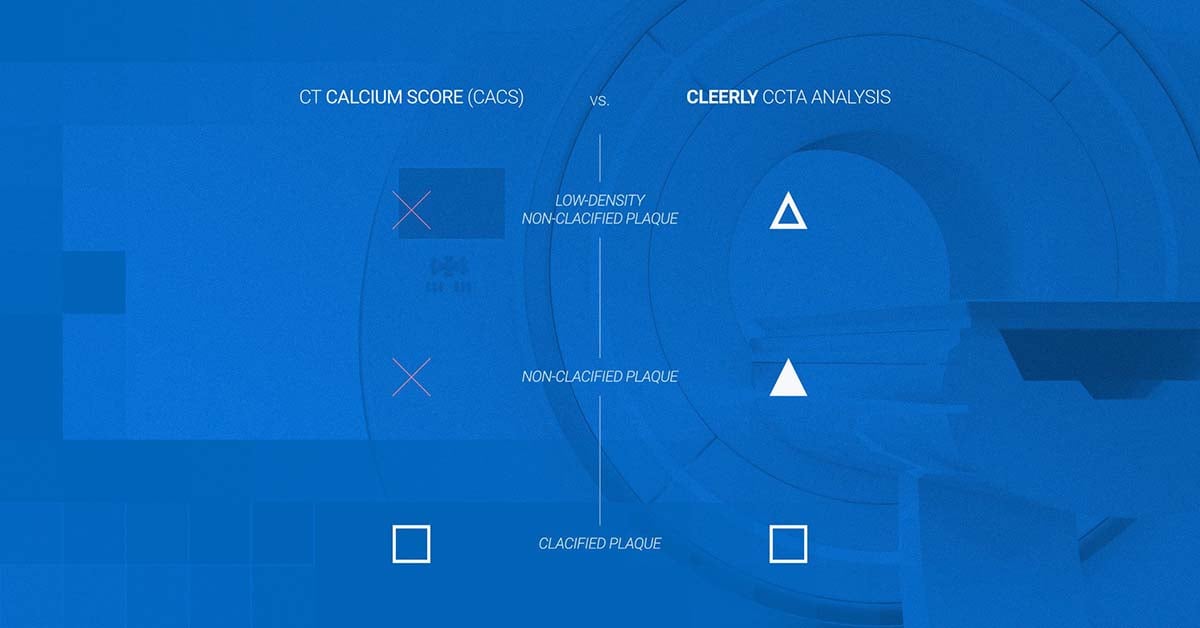
One of the most valuable data points is how much plaque buildup is in the coronary arteries. Coronary plaque is known as atherosclerosis and is a strong predictor of heart attack risk. Critically, a CCTA scan can distinguish between calcified plaque, which can cause a narrowing of the arteries, and low-density, non-calcified plaque, which can rupture and lead to a blood clot.
Additionally, a CCTA scan can measure ischemia, which is the restriction of blood flow arriving to the heart muscle, and also determine narrowing of the artery also known as a stenosis. Taken together, the measurements of ischemia, stenosis, and atherosclerosis can give a comprehensive picture of heart health.
The ability to apply analytics to CCTA scan results can provide cardiologists with further insights as well. Recently, the Innovations in Prevention Working Group of the American College of Cardiology introduced Atherosclerosis Treatment Algorithms, which provide recommendations for medical interventions based on CCTA screening results and traditional cardiovascular risk factors from existing clinical guidelines.3 These guidelines provide physicians with information that makes it possible to personalize treatment plans based on individual disease characteristics, assess the effectiveness of a given therapy, and change treatment plans over time.4
A traditional CT scan can detect calcium in coronary arteries. Cardiologists can calculate a coronary artery calcium score (CACS) from the percentage of hardened plaque in a patient’s arteries A CACS is a well-established measurement of coronary artery disease and heart attack risk, and recent research has shown it’s a better predictor than genetic risk.5
However, a CACS may not be a reliable measurement of heart health, as it only measures calcified plaque and can’t measure the riskier non-calcified plaque, stenosis or ischemia. Even when viewed in combination with additional markers of heart health such as hyperlipidemia (high cholesterol) or hypertension (high blood pressure), CACS is unable to provide a full picture of heart health.6
Multiple studies have shown that CCTA scan results are equivalent or superior to more traditional testing methodologies such as invasive coronary angiography and intravascular ultrasound (IVUS) – and without the need for a patient to undergo an invasive procedure.7,8,9 Ongoing or serialized use of CCTA scanning, similar to the practice of mammography for screening for breast cancer, shows promise as a way to measure CAD progression (and direct future treatment decisions) or regression (and demonstrate the effectiveness of medical therapy or lifestyle change).10
Meanwhile, increasing evidence shows that CCTA can be a valuable tool in treating CAD in addition to potentially diagnosing it. The SCOT HEART trial showed a 41% reduction in deaths and non-fatal heart attacks at a five-year endpoint among patients with stable chest pain who were treated with standard cardiovascular care plus CCTA, as compared to those treated with standard care alone.11
In addition, the DISCHARGE trial showed that patients managing CAD with standard care plus CCTA had a similar risk of major adverse cardiovascular events to patients managing CAD with standard care and ICA. However, the group treated with CCTA had a lower frequency of complication as well as revascularization, which is a procedure to restore blood flow to the arteries.12
In 2021, the American College of Cardiology and the American Heart Association updated their guidelines for the evaluation and diagnosis of chest pain, recommending CCTA as a “frontline testing strategy” for patients with stable and acute chest pain and no known history of CAD or intermediate to high risk of obstructive CAD. It’s estimated the new guidelines could apply to as many as 20 million patients in the United States.13,14
Cardiac CTA was given a Class 1, Level A recommendation, which is the strongest recommendation for a medical test (Class 1) and one that’s supported by evidence from multiple randomized clinical trials (Level A). Guidelines in the United States now align with guidelines published by the European Society of Cardiology in 2019.
A growing body of evidence shows that CCTA scans are equivalent or superior to many current standards for both diagnosing and treating CAD. What’s more, CCTA comes without the need for an invasive procedure and with the potential for serial scans that can be used to measure the progression or regression of heart disease over time. If it isn’t already, CCTA should be part of the standard of care for today’s cardiologists and their organizations.
References:
1. Armin Zadeh, MD, PhD. Coronary Computed Tomography Angiography (CCTA). Johns Hopkins Medicine. Accessed June 27, 2023. https://www.hopkinsmedicine.org/health/treatment-tests-and-therapies/coronary-computed-tomography-angiography-ccta ⇱
2. Nicolò Schicchi, Marco Fogante, Pierpaolo Palumbo, et al. The sub-millisievert era in CTCA: the technical basis of the new radiation dose approach. La radiologia medica. 2020 Nov;125(11):1024-1039. https://pubmed.ncbi.nlm.nih.gov/32930945/ ⇱
3. Andrew M. Freeman, MD; Subha V. Raman, MD; Monica Aggarwal, MD; et al. Integrating Coronary Atherosclerosis Burden and Progression with Coronary Artery Disease Risk Factors to Guide Therapeutic Decision Making. The American Journal of Medicine. 2023 March;136(3):260-269. https://www.sciencedirect.com/science/article/abs/pii/S0002934322008749 ⇱
4. Atherosclerosis Treatment Algorithms for Heart Disease Evaluation. Cleerly. 2023 February. https://cleerlyhealth.com/blog/atherosclerosis-treatment-algorithms ⇱
5. Sadiya S. Khan, MD, MSc; Wendy S. Post, MD; Xiuqing Guo, PhD; et al. Coronary artery calcium score and polygenic risk score for the prediction of coronary heart disease events. Journal of the American Medical Association. 2023;329(20):1768-1777. https://jamanetwork.com/journals/jama/article-abstract/2805138 ⇱
6. Cleerly Versus Coronary Artery Calcium Score: How the two heart disease tests measure up. Cleerly. 2023 June. https://cleerlyhealth.com/blog/cacs-versus-cleerly ⇱
7. Andrew D. Choi, Hugo Marques, Vishak Kumar, et al. CT Evaluation by Artificial Intelligence for Atherosclerosis, Stenosis and Vascular Morphology (CLARIFY): A Multi-center, international study. Journal of Cardiovascular Computed Tomography. 2021 June;15(6):470-476. https://www.journalofcardiovascularct.com/article/S1934-5925(21)00081-2/fulltext ⇱
8. William F. Griffin, Andrew D. Choi, Joanna S. Riess, et al. AI Evaluation of Stenosis on Coronary CTA, Comparison With Quantitative Coronary Angiography and Fractional Flow Reserve: A CREDENCE Trial Substudy. Journal of the American College of Cardiology: Cardiolovascular Imaging. 2023 Feb, 16 (2) 193–205. https://www.jacc.org/doi/10.1016/j.jcmg.2021.10.020 ⇱
9. Collin Fischer, Edward Hulten, Pallavi Belur, et al. Coronary CT angiography versus intravascular ultrasound for estimation of coronary stenosis and atherosclerotic plaque burden: a meta-analysis. JCTT. 2013 Jul-Aug;7(4):256-66. https://pubmed.ncbi.nlm.nih.gov/24148779/ ⇱
10. Dave Fornell. Cardiac CT soft plaque assessment may offer paradigm shift for coronary disease screening. Cardiovascular Business. July 2022. https://cardiovascularbusiness.com/topics/cardiac-imaging/computed-tomography-ct/cardiac-ct-soft-plaque-assessment-may-offer-paradigm ⇱
11. David Newby; Philip Adamson, MD; Colin Berry; et al. Coronary CT Angiography and 5-Year Risk of Myocardial Infarction. The New England Journal of Medicine. 2018 September; 379:924-933. https://www.nejm.org/doi/full/10.1056/NEJMoa1805971 ⇱
12. Pál Maurovich-Horvat, M.D., Ph.D., M.P.H.; Maria Bosserdt, Ph.D.; Klaus F. Kofoed, M.D.; CT or Invasive Coronary Angiography in Stable Chest Pain. NEJM. 2022; 386:1591-1602. https://www.nejm.org/doi/full/10.1056/NEJMoa2200963 ⇱
13. Martha Gulati; Phillip D. Levy; Debabrata Mukherjee; et al. 2021 AHA/ACC/ASE/CHEST/SAEM/SCCT/SCMR Guideline for the Evaluation and Diagnosis of Chest Pain: A Report of the American College of Cardiology/American Heart Association Joint Committee on Clinical Practice Guidelines. Circulation. 2021;144:368–454. https://www.ahajournals.org/doi/10.1161/CIR.0000000000001029 ⇱
14. Claire Johns. CCTA receives Multiple Class 1, Level A recommendations in 2021 New Chest Pain Guideline. Society of Cardiovascular Computed Tomography. 2021 October.
https://scct.org/news/585062/CCTA-receives-Multiple-Class-1-Level-A-recommendations-in-2021-New-Chest-Pain-Guideline-.htm ⇱

Sure, the taste of food is important, but do you know how your diet impacts your heart? Many foods contain hidden ingredients that negatively impact...

As we reach the end of 2023 and yet another remarkable year at Cleerly, it’s a great opportunity to reflect on all we’ve accomplished and begin...

Cleerly has had a busy few months – attending conferences, panel sessions, and winning awards as we all work together to achieve our mission of a...

Both coronary artery calcium score (CACS) and Cleerly evaluate heart disease risk using cardiac imaging. Learn about the key differences between the...

According to the American Heart Association (AHA), nearly half of Americans have some type of cardiovascular disease.1 Despite such wide prevalence,...

There's a little-known fact in cardiology: Most of the care that's provided doesn't prevent heart attacks.